43 diagram of a labeled microscope
Compound Microscope Parts - Labeled Diagram and their Functions - Rs ... Labeled diagram of a compound microscope Major structural parts of a compound microscope Optical components of a compound microscope Eyepiece Eyepiece tube Objective lenses Nosepiece Specimen stage Coarse and fine focus knobs Rack stop Illuminator Condenser Abbe condenser Iris Diaphragm Condenser Focus Knob Summary An overview of microscopes Labeling the Parts of the Microscope | Microscope activity, Science ... Description A collection of microscope diagrams and worksheets for science class. Download them all in one convenient PDF, and select the version that's best for your classroom. This PDF contains the following: 1. Parts of a Microscope Diagram - Color 2. Parts of a Microscope Diagram - Black and White 3.
Microscope labeled diagram - SlideShare Microscope labeled diagram Oct. 30, 2013 • 6 likes • 27,460 views Download Now Download to read offline Pisgah High School Follow 1. The Microscope Image courtesy of: Microscopehelp.com Basic rules to using the microscope 1. You should always carry a microscope with two hands, one on the arm and the other under the base. 2.

Diagram of a labeled microscope
A Study of the Microscope and its Functions With a Labeled Diagram These labeled microscope diagrams and the functions of its various parts, attempt to simplify the microscope for you. However, as the saying goes, 'practice makes perfect', here is a blank compound microscope diagram and blank electron microscope diagram to label. Download the diagrams and practice labeling the different parts of these ... Looking at the Structure of Cells in the Microscope When labeled with fluorescent dyes, they are invaluable for locating specific molecules in cells by fluorescence microscopy ... The confocal fluorescence microscope. This simplified diagram shows that the basic arrangement of optical components is similar to that of the standard fluorescence microscope shown in Figure 9-12, except that a laser is used to illuminate a … Compound Microscope Parts, Functions, and Labeled Diagram So, a compound microscope with a 10x eyepiece magnification looking through the 40x objective lens has a total magnification of 400x (10 x 40). Specimen or slide: The object used to hold the specimen in place along with slide covers for viewing. ... Compound Microscope Parts, Functions, and Labeled Diagram. Parts of a Compound Microscope.
Diagram of a labeled microscope. PDF Label parts of the Microscope: Answers Label parts of the Microscope: Answers Coarse Focus Fine Focus Eyepiece Arm Rack Stop Stage Clip . Created Date: 20150715115425Z ... Label the microscope — Science Learning Hub Label the microscope Add to collection Use this interactive to identify and label the main parts of a microscope. Drag and drop the text labels onto the microscope diagram. eye piece lens coarse focus adjustment high-power objective diaphragm or iris base fine focus adjustment light source stage Download Exercise Tweet Microscope Labeling - The Biology Corner Microscope Labeling. This simple worksheet pairs with a lesson on the light microscope, where beginning biology students learn the parts of the light microscope and the steps needed to focus a slide under high power. The labeling worksheet could be used as a quiz or as part of direct instruction where students label the microscope as you go ... Neuron under Microscope with Labeled Diagram - AnatomyLearner Here, you will also find the diagrams of different neuron types under a microscope. The neuron diagram shows the different parts (axon, dendrites, and cell body) of the neurons. You may also find more neuron diagrams on social media of anatomy learners. Frequently asked questions on a neuron under microscope
Label Microscope Diagram - EnchantedLearning.com Using the terms listed below, label the microscope diagram. arm - this attaches the eyepiece and body tube to the base. base - this supports the microscope. body tube - the tube that supports the eyepiece. coarse focus adjustment - a knob that makes large adjustments to the focus. diaphragm - an adjustable opening under the stage, allowing ... 22 Parts Of a Microscope With Their Function And Labeled Diagram The field diaphragm control is located around the lens located in the base. Hinge Screw -This screw fixes the arm to the base and allow for the tilting of the arm. Stage Clips - They hold the slide firmly onto the stage. On/OFF Switch - This switch on the base of the microscope turns the illuminator off and on. Parts of a microscope with functions and labeled diagram Figure: Diagram of parts of a microscope There are three structural parts of the microscope i.e. head, base, and arm. Head - This is also known as the body. It carries the optical parts in the upper part of the microscope. Base - It acts as microscopes support. It also carries microscopic illuminators. Parts of a Microscope Labeling Activity - Storyboard That Create a poster that labels the parts of a microscope and includes descriptions of what each part does. Click "Start Assignment". Use a landscape poster layout (large or small). Search for a diagram of a microscope. Using arrows and textables label each part of the microscope and describe its function.
Labeling the Parts of the Microscope Labeling the Parts of the Microscope This activity has been designed for use in homes and schools. Each microscope layout (both blank and the version with answers) are available as PDF downloads. You can view a more in-depth review of each part of the microscope here. Download the Label the Parts of the Microscope PDF printable version here. Compound Microscope Labeled Diagram | Quizlet QUESTION. The total magnification of a specimen being viewed with a 10X ocular lens and a 40X objective lens is. 15 answers. QUESTION. a mosquito beats its wings up and down 600 times per second, which you hear as a very annoying 600 Hz sound. if the air outside is 20 C, how far would a sound wave travel between wing beats. 2 answers. Microscope Diagram Labeled, Unlabeled and Blank | Parts of a Microscope ... This PDF contains the following: 1. Parts of a Microscope Diagram - Color 2. Parts of a Microscope Diagram - Black and White 3. Blank Parts of a Microscope Diagram - Black and White 4. Blank, Unlabeled Parts of a Microscope Diagram - Black and White 5. Blank Parts of a Microscope Diagram - Color 6. Blank, Unlabeled… Cat Skeleton Anatomy with Labeled Diagram - AnatomyLearner 29/05/2021 · The skeleton of all mammals, including the cat, supports the body and protects the deeper soft tissue structure. There are not so many differences found in the cat skeleton compare to the dog.In this article, I will discuss the osteological features of bones from cat skeleton anatomy. If you a beginner in learning veterinary anatomy, you may get confused to identify the …
MicroBichos: Saprolegnia sp. un organismo parasitoide
Virtual Microscope - NCBioNetwork.org Lesson Description BioNetwork’s Virtual Microscope is the first fully interactive 3D scope - it’s a great practice tool to prepare you for working in a science lab. Explore topics on usage, care, terminology and then interact with a fully functional, virtual microscope. When you are ready, challenge your knowledge in the testing section to see what you have learned.
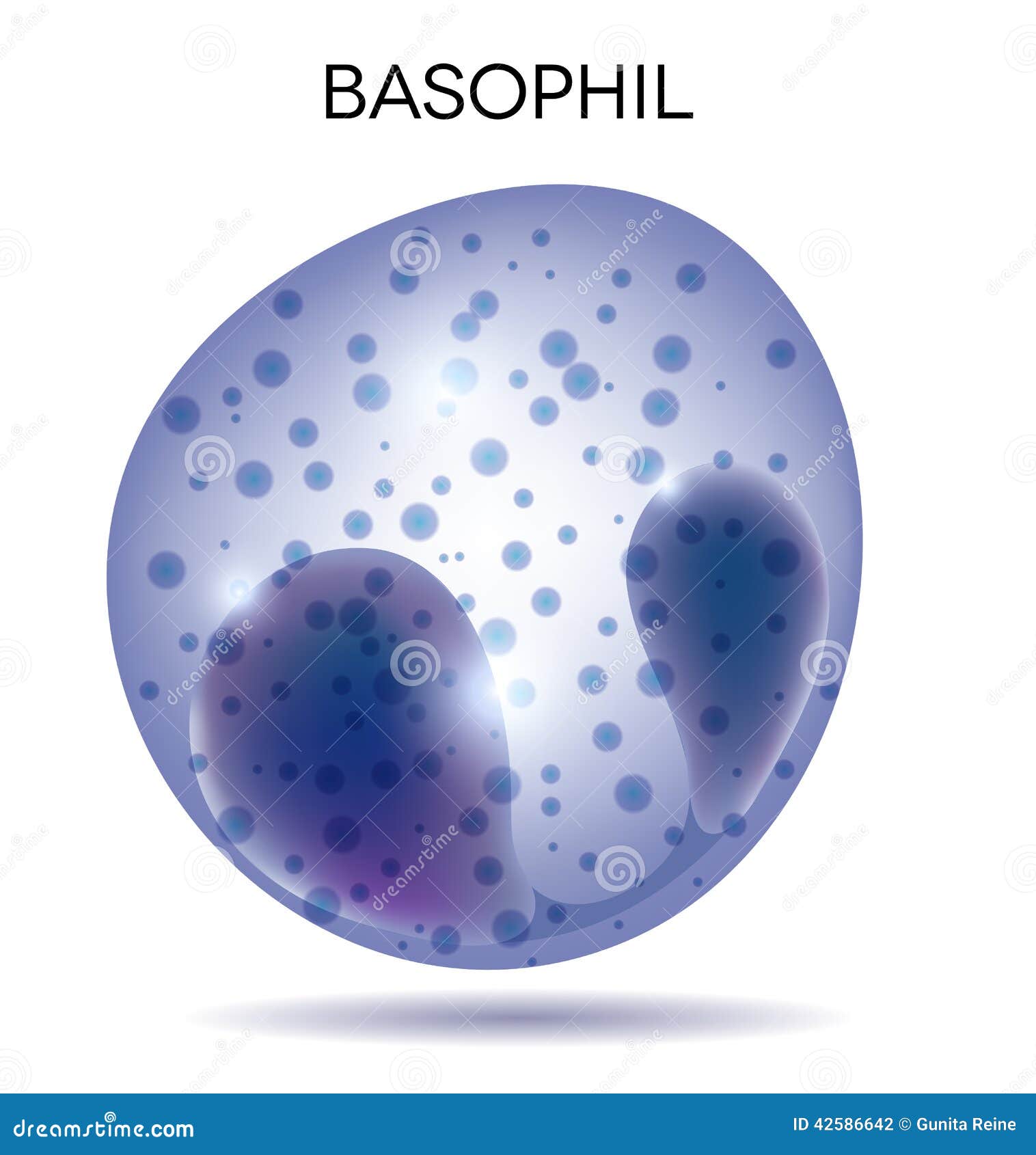
Basophil Cartoons, Illustrations & Vector Stock Images - 823 Pictures ...
Parts of the Microscope with Labeling (also Free Printouts) 5. Knobs (fine and coarse) By adjusting the knob, you can adjust the focus of the microscope. The majority of the microscope models today have the knobs mounted on the same part of the device. Image 5: The circled parts of the microscope are the fine and coarse adjustment knobs. Picture Source: bp.blogspot.com.

Roots, Stems and Leaves Diagrams
PDF Parts of a Microscope Printables - Homeschool Creations Label the parts of the microscope. You can use the word bank below to fill in the blanks or cut and paste the words at the bottom. Microscope Created by Jolanthe @ HomeschoolCreations.net. Parts of a eyepiece arm stageclips nosepiece focusing knobs illuminator stage objective lenses
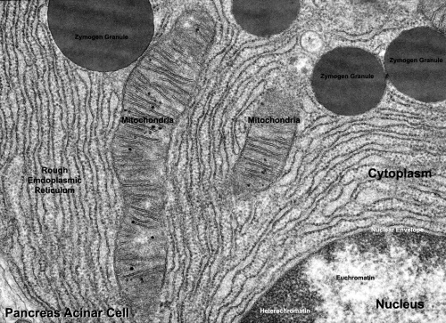
Talk:ANAT2511 Introduction to Histology - Embryology
Microscope, Microscope Parts, Labeled Diagram, and Functions The description given below summarize the brief description of microscope parts used to visualize the microscopic specimens such as animal cells, plant cells, microbes, bacteria, viruses, microorganisms etc. The Microscopes parts divided into three different structural parts Head, Base, and Arms. Head/Body: It contain the optical parts in the ...
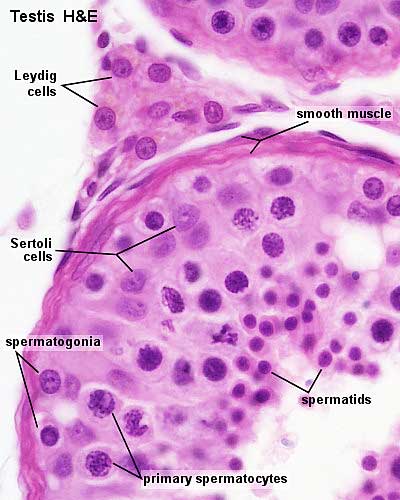
ANAT2341 Lab 1 - Spermatogenesis - Embryology
Label Microscope Diagram - EnchantedLearning.com Label Microscope Diagram Using the terms listed below, label the microscope diagram. Inventions and Inventors: arm - this attaches the eyepiece and body tube to the base. base - this supports the microscope. body tube - the tube that supports the eyepiece. coarse focus adjustment - a knob that makes large adjustments to the focus.
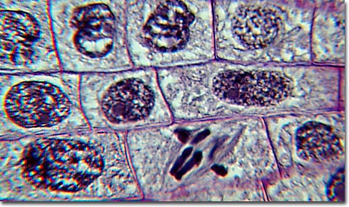
Molecular Expressions Photo Gallery: Mitosis
Labeled Microscope and Basics of Life Diagram | Quizlet PLAY. A microscope is an instrument widely to magnify and resolve the image of an object that is otherwise invisible to naked eye. For resolving the details of objects, which otherwise cannot be achieved by naked eye, a microscope is used. This set of flash cards will help the student to identify the different parts and function of the microscope.
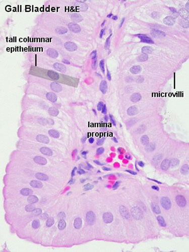
Gastrointestinal Tract - Gallbladder Histology - Embryology
Colon Histology Slide with Labeled Diagram - AnatomyLearner In addition, the colon labeled diagram also shows the bundle of nerve fibers (not seen under the binocular microscope). Finally, the labeled diagram shows a thin layer of tunica serosa that lines with a single layer of squamous cells. The mucosa of a colon labeled diagram. Let's see the second labeled diagram of the animal colon.

Lycopodium
Microscope Parts, Function, & Labeled Diagram - slidingmotion Microscope Parts Labeled Diagram The principle of the Microscope gives you an exact reason to use it. It works on the 3 principles. Magnification Resolving Power Numerical Aperture. Parts of Microscope Head Base Arm Eyepiece Lens Eyepiece Tube Objective Lenses Nose Piece Adjustment Knobs Stage Aperture Microscopic Illuminator Condenser Lens
Tissues Flashcards | Easy Notecards
Compound Microscope- Definition, Labeled Diagram, Principle, … 03/04/2022 · Therefore, a microscope can be understood as an instrument to observe tiny elements. The optical microscope often referred to as the light microscope, is a type of microscope that uses visible light and a system of lenses to magnify images of small subjects. There are two basic types of optical microscopes: Simple microscopes; Compound microscopes
Post a Comment for "43 diagram of a labeled microscope"