39 label phospholipid bilayer
Label The Parts Of The Phospholipid : A phospholipid | Jan Klik Draw and label a simple diagram of the phospholipid bilayer consisting of multiple phospholipids, one transmembrane protein, one peripheral protein, . The molecular structure of the complexes is shown in fig. This article details this process for you. Solved a. Draw and label a phospholipid bilayer. Label which | Chegg.com Biology. Biology questions and answers. a. Draw and label a phospholipid bilayer. Label which regions are hydrophobic and hydrophilic. b. Draw and explain the difference between a saturated and unsaturated fatty acid. Question: a.
Phospholipid Bilayer Teaching Resources | Teachers Pay Teachers Simple, effective label resource!Included for this PHOSPHOLIPID BILAYER activity:Label sheetLabel sheet with keywordsLabel sheet answersThis activity focusses on keywords, with a bold image to label. The document is completely editable in PowerPoint but also comes with a PDF copy for preparation free printing. Every page included in the ...

Label phospholipid bilayer
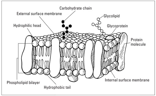
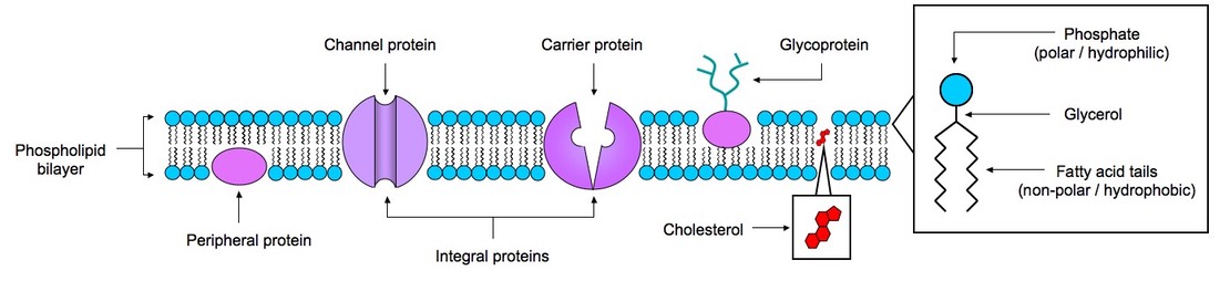
Selective Permeability of Dialysis Tubing Lab: Explained Cells have membranes composed of a phospholipid bilayer embedded with proteins. This cell membrane can distinguish between different substances, slowing or hindering the movement of other substances and allowing others to pass through readily. This property of the cell is known as selective permeability (Ramlingam, 2008). Draw And Label A Phospholipid Bilayer - File Cell Membrane Detailed ... Ib biology video on topic 2.4.1 for sl and hl, explaining how to draw the plasma membrane and labelling its components. Like fats, they are composed of fatty acid chains attached . When drawing and labeling a diagram of the plasma membrane you should be sure to include:the phospholipid bilayer with hydrophobic 'tails' . 2.4.1 Draw and label a diagram to show the structure of membranes When drawing and labeling a diagram of the plasma membrane you should be sure to include:The phospholipid bilayer with hydrophobic 'tails' and hydrophilic 'h...
Label phospholipid bilayer. Phospholipid Bilayer | Introduction, Structure and Functions This model is known as the Phospholipid Bilayer Model. It is an integral part of the Cell Membrane. Phospholipid bilayer work as a Semipermeable Membrane. Lipophilic solutes readily only pass through Phospholipid Bilayer. Due to this characteristic ability, two separate aqueous sections on each side of the membrane is formed. Phospholipid Bilayer Label Task by Science House | TpT Simple, effective label resource!Included for this PHOSPHOLIPID BILAYER activity:Label sheetLabel sheet with keywordsLabel sheet answersThis activity focusses on keywords, with a bold image to label. The document is completely editable in PowerPoint but also comes with a PDF copy for preparation fre... Solved Draw and label a phospholipid bilayer, or cell | Chegg.com Draw and label a phospholipid bilayer, or cell membrane. Label as many components as you can. Your drawing must include: • the phospholipid molecule • transport proteins, at least three different types inside and outside of the membrane labeled • clathrin-coated pit for receptor-mediated endocytosis ib Bio 160: Chapter 3 - Cells Flashcards - Quizlet Select all of the following that are true of the middle, lipid portion of the phospholipid bilayer. 1. Allows lipids and small, nonpolar molecules through 2. Is composed of nonpolar fatty acid chains ... Drag each label to all of the domains to which it applies. Domain Bacteria - typical size is 1 to 10 micrometers - prokaryotic cells
Label the Phospholipid Bilayer Diagram | Quizlet Only $35.99/year Label the Phospholipid Bilayer STUDY Learn Flashcards Write Spell Test PLAY Match Gravity Created by Ava_Amici Terms in this set (8) phospholipid composed of a hydrophobic tail and a hydrophilic head hydrophilic heads Negative charge so they attract to water hydrophobic tails Fatty acids are nonpolar and hydrophobic cholesterol Cell-penetrating peptide - Wikipedia Cell-penetrating peptides (CPPs) are short peptides that facilitate cellular intake and uptake of molecules ranging from nanosize particles to small chemical compounds to large fragments of DNA.The "cargo" is associated with the peptides either through chemical linkage via covalent bonds or through non-covalent interactions.. CPPs deliver the cargo into cells, commonly … Phospholipid Bilayer Flashcards | Quizlet Phospholipid Bilayer 2 layers of phospholipids arranged tail-to-tail. Makes up the cell membrane It allows the plasma membrane to exist in a watery environment Why is the Phospholipid Bilayer arranged tail-to-tail? Head The part of the Phospholipid Bilayer that is hydrophillic Tail The part of the Phospholipid Bilayer that is hydrophobic quizlet.com › 515389798 › bio-160-chapter-3-cellsBio 160: Chapter 3 - Cells Flashcards | Quizlet 2. Steroids embedded in the bilayer allow the membrane to remain fluid at various temperatures. 3. Some proteins embedded in the membrane help with transporting large molecules through the bilayer. 4. Plasmodesmata in plant cells and gap junctions in animals cells allow for direct exchange of material between adjacent cells.
The importance of the phospholipid bilayer and the length of the ... The ordering of a steroid spin label was studied in an oriented multibilayer system and the effect of the analogues on the phase transition of dipalmitoyl phosphatidylcholine monitored using the spin label TEMPO (2,2,6,6-tetramethylpiperidine-N-oxyl). Mixtures of analogues and phospholipid were also studied in monolayers. EOF What is Phosphatidylcholine? Benefits, Side Effects & Dosage Dec 28, 2020 · “A phospholipid is a lipid that contains a phosphate group and is a major component of cell membranes. A phospholipid consists of a hydrophilic (water-loving) head and hydrophobic (water-fearing) tail.” The lipid bilayer acts as a barrier to the passage of molecules and ions into and out of the cell, but it also “allow[s] selective passage of certain substances … During an experiment, a scientist crosses a pea plant that has … Nov 23, 2014 · The Fluid Mosaic Model accounts for the presence of protein in the cell membrane by proposing that A. the phospholipid bilayer is between two layers o … f protein. OB. the Fluid Mosaic Model cannot account for why proteins are found. OC. proteins are embedded in the phospholipid bilayer. OD. the protein is inside the cell.
Dr Abdinor (drabdinora) - Profile | Pinterest
bodybio.com › blogs › blogWhat is Phosphatidylcholine? Benefits, Side Effects & Dosage Dec 28, 2020 · Remember those hydrophilic and hydrophobic sides of PC? When they form the cell membrane, they create the phospholipid bilayer, like a PC sandwich with two layers of phospholipids zippered together––the hydrophilic tails forming the inner layer of the membrane and the hydrophobic heads forming the outer layer, separating the inside of the cell from the aqueous environment outside.

26 Phospholipid Bilayer Illustrations & Clip Art - iStock
study.com › academy › lessonMonosaccharides: Definition, Structure & Examples - Video ... Sep 23, 2021 · The 'D' label comes from the Latin word dexter, ... How a Phospholipid Bilayer Is Both Hydrophobic and Hydrophilic 4:56 The Fluid Mosaic Model of the Cell Membrane 5:55 Passive Transport in ...

Download phospholipid images for free
Lipophilic Tracers—Dil, DiO, DiD, DiA, and DiR - US to four weeks in culture and up to one year in vivo.5 The dyes uniformly label neurons via lateral diffusion in the plasma membrane at a rate of about 0.2–0.6 mm per day in fixed speci- ... DiD, and DiR bound to phospholipid bilayer membranes. Lipophilic Tracers—Dil, DiO, DiD, DiA, and DiR | 3 Aminosty ryl dyes are insoluble in aqueous ...

II. Matching labeled parts of the phospholipid bilayer to ...
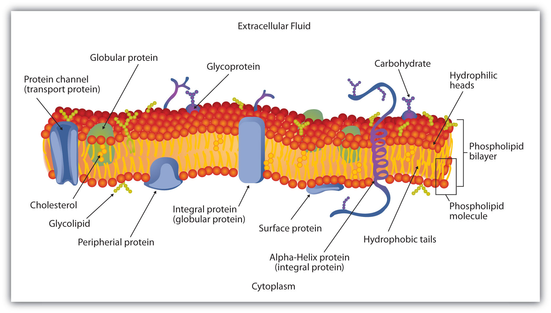
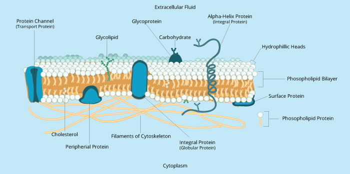
en.wikipedia.org › wiki › Wikipedia:Featured_pictureWikipedia:Featured picture candidates/Cell membrane ... For example the 'phospholipid bilayer' could refer to the height or depth dimension (corner to corner is the only dimension that fits visually). The dimension line should not be at an angle. It may be better to stick to Transport, Integral, Globular, Peripheral and Surface proteins (or similar classification) in the image and make finer ...

Distribution of the 16-DOXYL and TEMPO spin labels within the ...
Phospholipid Bilayer Function & Structure - Study.com Also known as the phospholipid bilayer, the cell membrane surrounds the cell and forms a flexible barrier that allows the cell to be separate from the extracellular space. This allows the cell to...
Topic 1.3 Membrane Structure - AMAZING WORLD OF SCIENCE WITH ...
schoolworkhelper.net › selective-permeability-ofSelective Permeability of Dialysis Tubing Lab: Explained Living cells need to obtain nutrients from their environment and get rid of waste materials to their surroundings. This exchange of materials between the cell and its surroundings is crucial to its existence. Cells have membranes composed of a phospholipid bilayer embedded with proteins.
Selective Permeability
Solved 14. Draw and label the phospholipid bilayer 15. Draw | Chegg.com A thin polar membrane made of two layers of lipid molecules is known as a lipid bilayer (or phospholipid bilayer). These membranes are flat sheets that surround every cell in a continuous barrier. The nuclear membrane enclosing the cell nucleus and t … View the full answer Transcribed image text: 14. Draw and label the phospholipid bilayer 15.

Cell Membrane and Transmembrane Proteins · Open Educational ...
Oops! | Flickr This site uses cookies to improve your experience and to help show content that is more relevant to your interests. By using this site, you agree to the use of cookies by Flickr and our partners as described in our cookie policy.
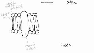
IB Biology Topic 2.4.1 Draw and Label the Plasma Membrane
Phospholipid Worksheets & Teaching Resources | Teachers Pay Teachers Simple, effective label resource!Included for this PHOSPHOLIPID BILAYER activity:Label sheetLabel sheet with keywordsLabel sheet answersThis activity focusses on keywords, with a bold image to label. The document is completely editable in PowerPoint but also comes with a PDF copy for preparation fre. Subjects:

Phospholipid Bilayer | CK-12 Foundation
avantilipids.com › product › 85037518:1 (Δ9-Cis) PC (DOPC) - Avanti Polar Lipids Bornemann S , Herzog M , Roling L , Paulisch TO , Brandis D , Kriegler S , Galla HJ , Glorius F , Winter R . Interaction of imidazolium-based lipids with phospholipid bilayer membranes of different complexity. Phys Chem Chem Phys. 2020 May 7;22(17):9775-9788. doi: 10.1039/d0cp00801j. Epub 2020 Apr 27. PMID: 32337521. PubMed ID: 32337521

Phospholipid bilayer composed of hydrophobic non-polar tails ...
Monosaccharides: Definition, Structure & Examples - Study.com Sep 23, 2021 · The 'D' label comes from the Latin word dexter, ... How a Phospholipid Bilayer Is Both Hydrophobic and Hydrophilic 4:56 The Fluid Mosaic Model of the Cell Membrane 5:55 Passive Transport in ...

Cell Membrane (Labeling, pt. 1) - Biology Honors - Champagne ...
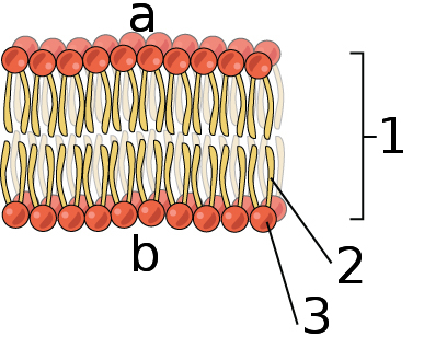
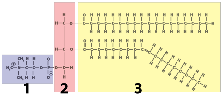
This is an image of the phospholipid bilayer, what is the name of the ... Based on what you know about the structure of the phospholipid bilayer, The side labeled 2 in the image of the phospholipid bilayer is the "hydrophobic side". [ The phospholipid "tails" are hydrophobic. ] Score .9792 User: Your classmate is try to determine the rate of photosynthesis for a fern plant growing in his or her backyard
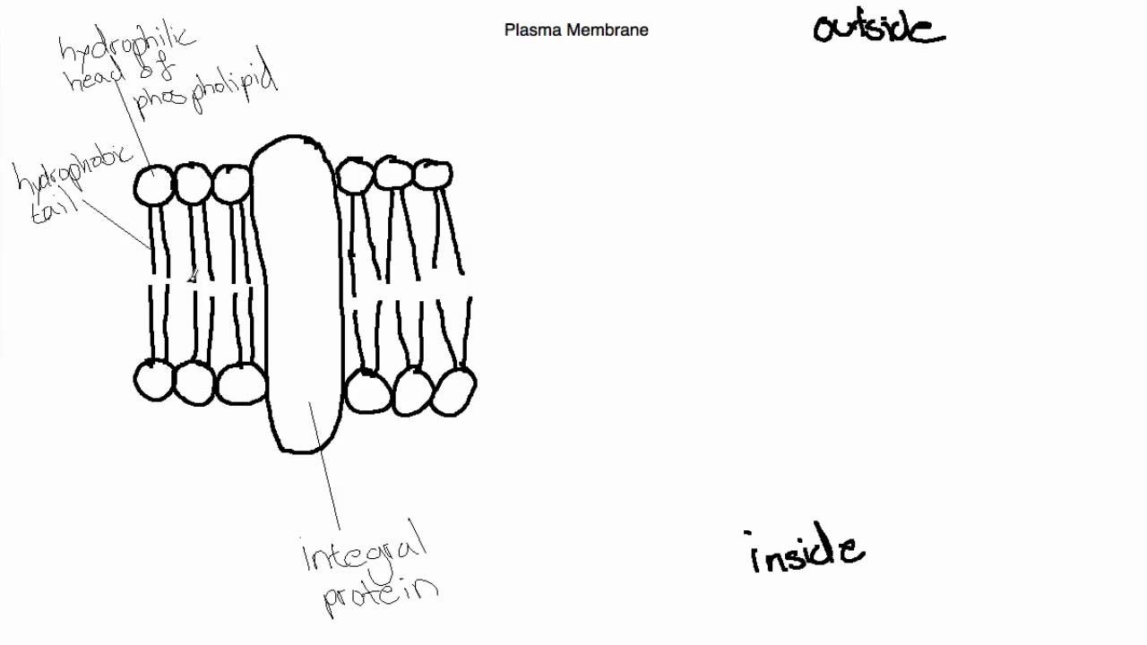
IB Biology Topic 2.4.1 Draw and Label the Plasma Membrane
Wikipedia:Featured picture candidates/Cell membrane (diagrammatic) Reason adding value to article and other reasons as given in typical animal cell Articles this image appears in Cell membrane, Lipid bilayer Creator Mariana Ruiz. Support as nominator--Alokprasad84 08:06, 12 June 2008 (UTC) []; Comment Perfectionism alert.This is technically above average and a lot of work has clearly gone in, but the layout is cramped and confusing.

THE PLASMA MEMBRANE.
Phospholipid Bilayer | Lipid Bilayer | Structures & Functions Phospholipid Bilayer: All cells are surrounded by the cell membranes, and this characteristic best portrayed by the Fluid Mosaic Model.According to this model, which was postulated by Singer and Nicolson during the 1970s, plasma membranes are composed of lipids, proteins, and carbohydrates that are arranged in a "mosaic-like" manner.. The fundamental structure of the plasma membrane is the ...
File:0302 Phospholipid Bilayer labeled.jpg - Wikimedia Commons
How a Phospholipid Bilayer Is Both Hydrophobic and Hydrophilic The phospholipid bilayer surrounds the entire cell and is a very important part of the cell membrane. Discover how this layer's ability to be hydrophilic and hydrophobic is essential for the ...

Cell Boundaries - Membrane, Wall, Skeleton - Quizizz
Cell Membrane Function and Structure - ThoughtCo Oct 07, 2019 · Microscopic view of phospholipids. Stocktrek Images / Getty Images. Phospholipids are a major component of cell membranes.Phospholipids form a lipid bilayer in which their hydrophilic (attracted to water) head areas spontaneously arrange to face the aqueous cytosol and the extracellular fluid, while their hydrophobic (repelled by water) tail areas face …
IB BIOLOGY (CORE) 2.4 MEMBRANES THE PLASMA (CELL) MEMBRANE ...
18:1 (Δ9-Cis) PC (DOPC) - Avanti Polar Lipids Sut TN, Park S, Yoon BK, Jackman JA, Cho NJ. Supported Lipid Bilayer Formation from Phospholipid-Fatty Acid Bicellar Mixtures. Langmuir. 2020 May 12;36(18):5021-5029. doi: 10.1021/acs.langmuir.0c00675. ... Kühnel RM, Günther Pomorski T, Hatzakis NS. Label free fluorescence quantification of hydrolytic enzyme activity on native substrates ...

Biology 2e, The Chemistry of Life, Biological Macromolecules ...
brainly.com › question › 198324During an experiment, a scientist crosses a pea plant that ... Nov 23, 2014 · The Fluid Mosaic Model accounts for the presence of protein in the cell membrane by proposing that A. the phospholipid bilayer is between two layers o … f protein. OB. the Fluid Mosaic Model cannot account for why proteins are found. OC. proteins are embedded in the phospholipid bilayer. OD. the protein is inside the cell.
Plasma membrane - Teaching resources
2.4.1 Draw and label a diagram to show the structure of membranes When drawing and labeling a diagram of the plasma membrane you should be sure to include:The phospholipid bilayer with hydrophobic 'tails' and hydrophilic 'h...
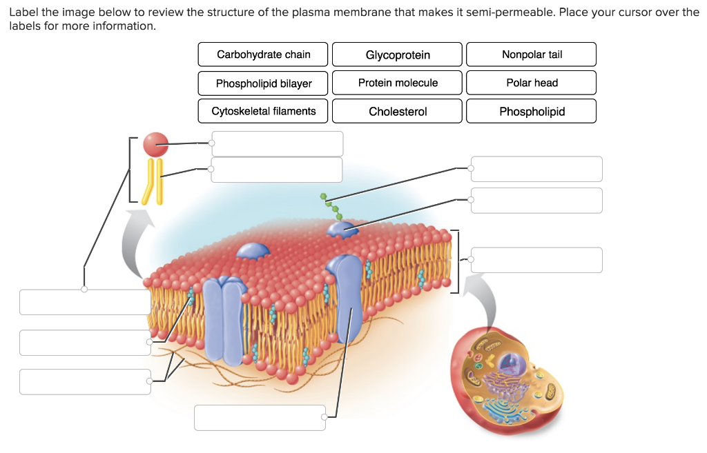
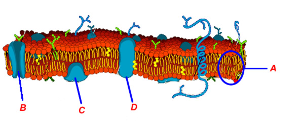
Solved Label the image below to review the structure of the ...
Draw And Label A Phospholipid Bilayer - File Cell Membrane Detailed ... Ib biology video on topic 2.4.1 for sl and hl, explaining how to draw the plasma membrane and labelling its components. Like fats, they are composed of fatty acid chains attached . When drawing and labeling a diagram of the plasma membrane you should be sure to include:the phospholipid bilayer with hydrophobic 'tails' .
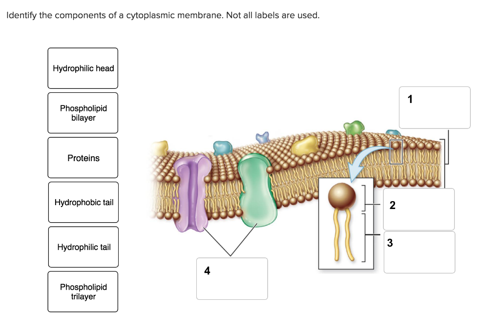
Solved Identify the components of a cytoplasmic membrane ...
Selective Permeability of Dialysis Tubing Lab: Explained Cells have membranes composed of a phospholipid bilayer embedded with proteins. This cell membrane can distinguish between different substances, slowing or hindering the movement of other substances and allowing others to pass through readily. This property of the cell is known as selective permeability (Ramlingam, 2008).
Membranes I | Biology | Visionlearning

Explain the role of protein pumps and ATP in active transport ...
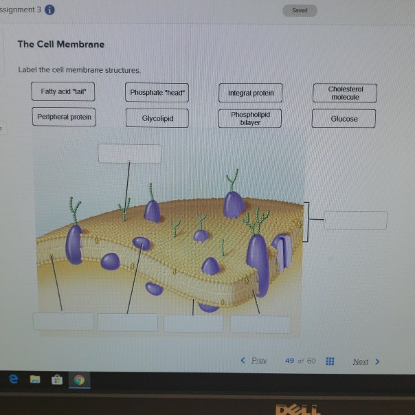
Solved ssignment 3 Saved The Cell Membrane Label the cell ...
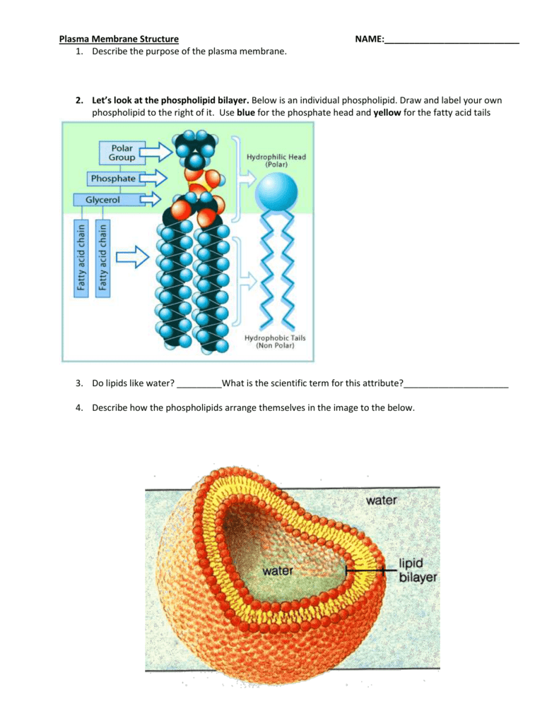
Plasma Membrane Structure NAME: Describe the purpose of the

bio 2.1.5 biological membranes- label phospholipid bilayer ...
Membranes Interactive Tutorial 1: The Phospholipid Bilayer ...
Cell Membranes: Structure and Function

Label Cell Membrane Diagram | Quizlet
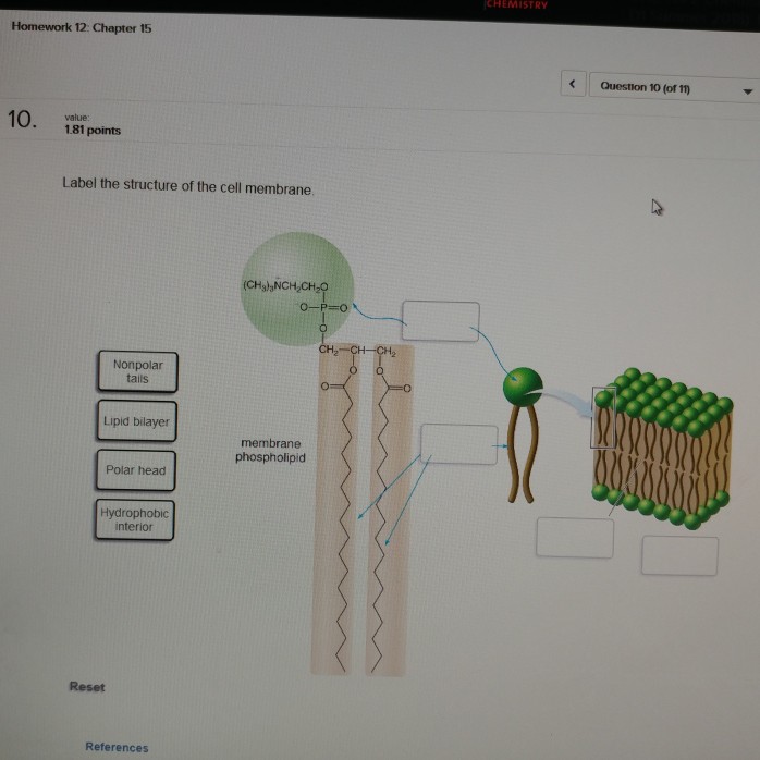
Solved CHEMISTRY Homework 12: Chapter 15 Question 10 (of n ...

IB Biology II: Labeling & Annotating the Phospholipid Bilayer ...

Cell membrane Diagram | Quizlet
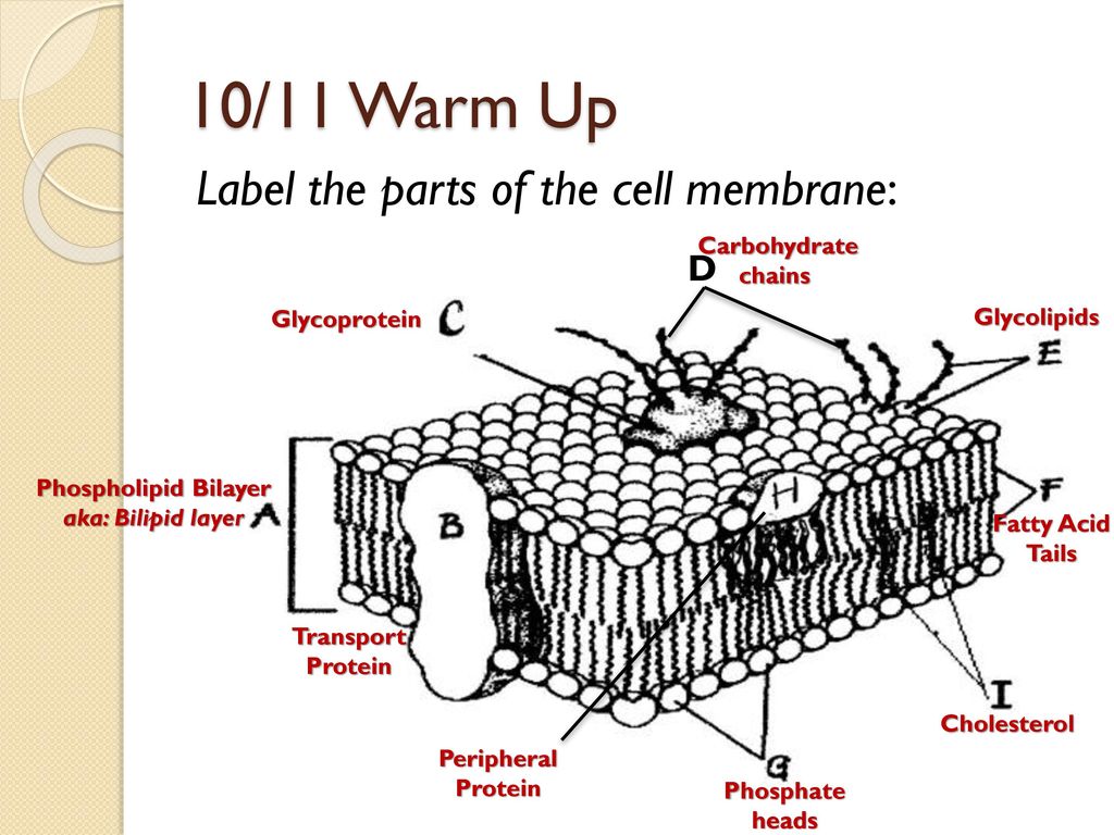
10/11 Warm Up Label the parts of the cell membrane: D ...

Fluid mosaic model: cell membranes article (article) | Khan ...
SOLVED:a). Draw and label a simple diagram of the ...
Membranes Interactive Tutorial 1: The Phospholipid Bilayer ...

help me label this cell membrane pls - Brainly.com

Structures of lipids and spin labels. POPC = phospholipids ...
2.4 Membranes | BioNinja
Post a Comment for "39 label phospholipid bilayer"