45 diagram of microscope with label
A Study of the Microscope and its Functions With a Labeled Diagram May 21, 2019 - To better understand the structure and function of a microscope, we need to take a look at the labeled microscope diagrams of the compound ... Microscope Diagram Labeled, Unlabeled and Blank | Parts of a ... Print a microscope diagram, microscope worksheet, or practice microscope quiz in ... In addition to labeling the microscope parts, students are asked to ...
Sperm Under Microscope with Labeled Diagram - AnatomyLearner Structure of sperm under a microscope The head of a sperm The neck of the spermatozoa The middle piece of the spermatozoa The principal piece of the sperm's tail The end piece of the sperm's tail Sperm in the epididymis Cyclic events in the seminiferous tubules Sperm under microscope 400x labeled Sperm with 40x and 100x labeled diagram

Diagram of microscope with label
Parts of the Microscope with Labeling (also Free Printouts) Parts of the Microscope with Labeling (also Free Printouts) By Editorial Team March 7, 2022 A microscope is one of the invaluable tools in the laboratory setting. It is used to observe things that cannot be seen by the naked eye. Table of Contents 1. Eyepiece 2. Body tube/Head 3. Turret/Nose piece 4. Objective lenses 5. Knobs (fine and coarse) 6. Compound Microscope - Diagram (Parts labelled), Principle and Uses Image : Labeled Diagram of compound microscope parts See: Labeled Diagram showing differences between compound and simple microscope parts Structural Components The three structural components include 1. Head This is the upper part of the microscope that houses the optical parts 2. Arm Labeling the Parts of the Microscope | Microscope World Resources Microscope World explains the parts of the microscope, including a printable worksheet for schools and home. Need Asssistance? 800-942-0528. Microscope Blog ... Labeling the Parts of the Microscope. This activity has been designed for use in homes and schools. Each microscope layout (both blank and the version with answers) are available as PDF ...
Diagram of microscope with label. Microscope Labeling Game - PurposeGames.com Microscope Labeling Game — Quiz Information. This is an online quiz called Microscope Labeling Game. There is a printable worksheet available for download here so you can take the quiz with pen and paper. This quiz is filed in the following categories. Science. › cells › bactcellInteractive Bacteria Cell Model - CELLS alive Ribosomes: Ribosomes give the cytoplasm of bacteria a granular appearance in electron micrographs.Though smaller than the ribosomes in eukaryotic cells, these inclusions have a similar function in translating the genetic message in messenger RNA into the production of peptide sequences (proteins). Microscope Parts and Functions It also allows the specimen to be labeled, transported, and stored without damage. Stage: The flat platform where the slide is placed. Stage clips: Metal clips that hold the slide in place. Stage height adjustment (Stage Control): These knobs move the stage left and right or up and down. Microscope With Labels Clip Art at Clker.com PEOPLE GOT HERE BY SEARCHING: diagrams of the microscope · light microscope and label · the compound microscope drawing · diagram of microscope with labelling ...
Compound Microscope Parts – Labeled Diagram and their Functions What is a "compound microscope"? Labeled diagram of a compound microscope Major structural parts of a compound microscope Optical components of a compound microscope Eyepiece Eyepiece tube Objective lenses Nosepiece Specimen stage Coarse and fine focus knobs Rack stop Illuminator Condenser Abbe condenser Iris Diaphragm Condenser Focus Knob Summary microbenotes.com › parts-of-a-microscopeParts of a microscope with functions and labeled diagram Sep 17, 2022 · Figure: Diagram of parts of a microscope. There are three structural parts of the microscope i.e. head, base, and arm. Head – This is also known as the body. It carries the optical parts in the upper part of the microscope. Base – It acts as microscopes support. It also carries microscopic illuminators. Simple Microscope - Diagram (Parts labelled), Principle, Formula ... A simple microscope consists of Optical parts Mechanical parts Labeled Diagram of simple microscope parts Optical parts The optical parts of a simple microscope include Lens Mirror Eyepiece Lens A simple microscope uses biconvex lens to magnify the image of a specimen under focus. alive! Since 1994, CELLS alive! has provided students with a learning resource for cell biology, microbiology, immunology, and microscopy through the use of mobile-friendly interactive animations, video, puzzles, quizzes and study aids.
Microscope With Labels clip art - Pinterest Microscope Diagram Labeled, Unlabeled and Blank | Parts of a Microscope. Print a microscope diagram, microscope worksheet, or practice microscope quiz in order ... Compound Microscope: Definition, Diagram, Parts, Uses, Working ... - BYJUS Compound microscope is a type of optical microscope that is used for obtaining a high-resolution image. There are more than two lenses in a compound microscope. Learn about the working principle, parts and uses of a compound microscope along with a labeled diagram here. A Study of the Microscope and its Functions With a Labeled Diagram ... A Study of the Microscope and its Functions With a Labeled Diagram To better understand the structure and function of a microscope, we need to take a look at the labeled microscope diagrams of the compound and electron microscope. These diagrams clearly explain the functioning of the microscopes along with their respective parts. › 2022/09/16 › governor-newsom-signsGovernor Newsom Signs Sweeping Climate Measures, Ushering in ... Sep 16, 2022 · New California laws will create 4 million jobs, reduce the state’s oil use by 91%, cut air pollution by 60%, protect communities from oil drilling, and accelerate the state’s transition to clean…
Diagram of a Microscope by ScienceDoodles on DeviantArt
Labelled Diagram of Compound Microscope The below mentioned article provides a labelled diagram of compound microscope. Part # 1. The Stand: The stand is made up of a heavy foot which carries a curved inclinable limb or arm bearing the body tube. The foot is generally horse shoe-shaped structure (Fig. 2) which rests on table top or any other surface on which the microscope in kept.

Compound Microscope Parts
Microscope Labeling - The Biology Corner Students label the parts of the microscope in this photo of a basic laboratory light microscope. Can be used for practice or as a quiz. ... Microscope Labeling . Microscope Use: 15. When focusing a specimen, you should always start with the _____ objective. 16. When using the high power objective, only the _____ knob should be used. 17. The ...
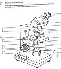
Answered: Microscope Structure and Function… | bartleby
Microscope Parts, Function, & Labeled Diagram - slidingmotion Microscope parts labeled diagram gives us all the information about its parts and their position in the microscope. Microscope Parts Labeled Diagram The principle of the Microscope gives you an exact reason to use it. It works on the 3 principles. Magnification Resolving Power Numerical Aperture. Parts of Microscope Head Base Arm Eyepiece Lens
Label a microscope - Teaching resources
Microscope Labeling Diagram | Quizlet Microscope Labeling 4.2 (62 reviews) + − Flashcards Learn Test Match Created by Terms in this set (12) Body Tube Separates the eyepiece lens from the objective lenses. Eyepiece Contains the ocular lens and magnifies the image produced by the objective lenses. Coarse Focus Knob Moves the stage large distances to roughly focus the image.

Parts of a Microscope Labeling Activity
› 6-label-the-microscopeLabel the microscope — Science Learning Hub Jun 08, 2018 · All microscopes share features in common. In this interactive, you can label the different parts of a microscope. Use this with the Microscope parts activity to help students identify and label the main parts of a microscope and then describe their functions. Drag and drop the text labels onto the microscope diagram. If you want to redo an ...

Draw a neat labelled diagram of a compound microscope. Derive ...
› lifestyleLifestyle | Daily Life | News | The Sydney Morning Herald The latest Lifestyle | Daily Life news, tips, opinion and advice from The Sydney Morning Herald covering life and relationships, beauty, fashion, health & wellbeing

File:Labelledmicroscope.gif - Wikimedia Commons
Labeling the Parts of the Microscope Labeling the Parts of the Microscope This activity has been designed for use in homes and schools. Each microscope layout (both blank and the version with answers) are available as PDF downloads. You can view a more in-depth review of each part of the microscope here. Download the Label the Parts of the Microscope PDF printable version here.

Microscope Parts and Functions
Microscope, Microscope Parts, Labeled Diagram, and Functions Microscope, Microscope Parts, Labeled Diagram, and Functions What is Microscope? A microscope is a laboratory instrument used to examine objects that are too small to be seen by the naked eye. It is derived from Ancient Greek words and composed of mikrós, "small" and skopeîn,"to look" or "see".
Microscope Parts & Specifications | Microscope World Resources
Label Microscope Diagram - EnchantedLearning.com Label Microscope Diagram Using the terms listed below, label the microscope diagram. Inventions and Inventors arm - this attaches the eyepiece and body tube to the base. base - this supports the microscope. body tube - the tube that supports the eyepiece. coarse focus adjustment - a knob that makes large adjustments to the focus.

Compound Microscope Parts – Labeled Diagram and their ...
Microscope Diagram Labeled, Unlabeled and Blank - Pinterest Oct 6, 2019 - Print a microscope diagram, microscope worksheet, or practice microscope quiz in order to learn all the parts of a microscope. Pinterest. ... Study guide for the digestive system focusing on vocabulary and labeling diagrams; intended for high school students taking anatomy and physiology. Geneé Handley. Anatomy - Digestive System.

Labelling a Microscope Diagram | Quizlet
Diagram of a Compound Microscope - Biology Discussion Information recorded on adhesive label is stuck to the base of the microscope for future reference. (ii) Use: Having calibrated the eyepiece scale for all the ...

Simple Microscope - Parts, Functions, Diagram and Labelling ...
Label the Microscope Diagram | Download Scientific Diagram - ResearchGate Download scientific diagram | Label the Microscope Diagram from publication: Laboratory Exercises in Microbiology: Discovering the Unseen World through Hands-on Investigation | Microbiology ...

Parts of a Microscope with Their Functions – Microbe Online
rsscience.com › stereo-microscopeParts of Stereo Microscope (Dissecting microscope) – labeled ... Labeled part diagram of a stereo microscope Major structural parts of a stereo microscope. There are three major structural parts of a stereo microscope. The viewing Head includes the upper part of the microscope, which houses the most critical optical components, including the eyepiece, objective lens, and light source of the microscope.

Parts of Stereo Microscope (Dissecting microscope) – labeled ...
Microscope Labeling - The Biology Corner 1) Start with scanning (the shortest objective) and only use the COARSE knob . Once it is focused… 2) Switch to low power (medium) and only use the COARSE knob . You may need to recenter your slide. Once it is focused.. 3) Switch to high power (long objective).

Microscope With Labels clip art | Microscope parts, Science ...
Microscope Diagram Labeled, Unlabeled and Blank - Pinterest Microscope Diagram Labeled, Unlabeled and Blank | Parts of a Microscope - Tim's Printables. Print a microscope diagram, microscope worksheet, or practice microscope quiz in order to learn all the parts of a microscope. Tim's Printables. 40k followers . Life Science Middle School ...

Different types of Microscopes – light microscope, electron ...
PDF Label parts of the Microscope Label parts of the Microscope: . Created Date: 20150715115425Z

Compound Microscope Parts, Functions, and Labeled Diagram ...
Microscope labeled diagram - SlideShare 1 of 2 Microscope labeled diagram Oct. 30, 2013 • 6 likes • 28,252 views Download Now Download to read offline Pisgah High School Follow Advertisement Recommended Microscope Basics Mrs. Henley 3.4k views • 7 slides Parts and Functions of the Compound Microscope IsaganiDioneda 3.3k views • 43 slides SCIENCE7: The Microscope Christian Adriano-ursabia

Compound Microscope Parts – Labeled Diagram and their ...
16 Parts of a Compound Microscope: Diagrams and Video Once you have an understanding of the parts of the microscope it will be much easier to navigate around and begin observing your specimen, which is the fun part! The 16 core parts of a compound microscope are: Head (Body) Arm. Base. Eyepiece. Eyepiece tube.

Compound Microscope Parts, Diagram Definition, Application ...
Compound Microscope Parts, Functions, and Labeled Diagram Compound Microscope Definitions for Labels Eyepiece (ocular lens) with or without Pointer: The part that is looked through at the top of the compound microscope. Eyepieces typically have a magnification between 5x & 30x. Monocular or Binocular Head: Structural support that holds & connects the eyepieces to the objective lenses.
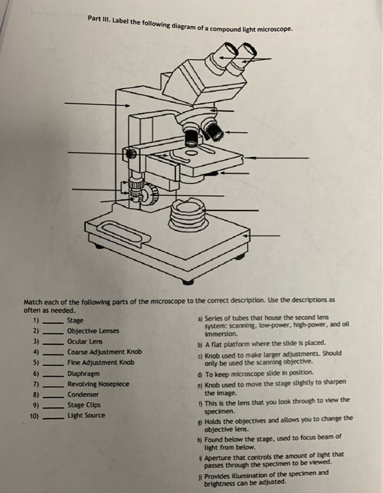
Solved Part III. Label the following diagram of a compound ...
Labeling the Parts of the Microscope | Microscope World Resources Microscope World explains the parts of the microscope, including a printable worksheet for schools and home. Need Asssistance? 800-942-0528. Microscope Blog ... Labeling the Parts of the Microscope. This activity has been designed for use in homes and schools. Each microscope layout (both blank and the version with answers) are available as PDF ...

Diagram of traveling microscope setup with implant cast and ...
Compound Microscope - Diagram (Parts labelled), Principle and Uses Image : Labeled Diagram of compound microscope parts See: Labeled Diagram showing differences between compound and simple microscope parts Structural Components The three structural components include 1. Head This is the upper part of the microscope that houses the optical parts 2. Arm

Microscope Diagram Labeled, Unlabeled and Blank | Parts of a ...
Parts of the Microscope with Labeling (also Free Printouts) Parts of the Microscope with Labeling (also Free Printouts) By Editorial Team March 7, 2022 A microscope is one of the invaluable tools in the laboratory setting. It is used to observe things that cannot be seen by the naked eye. Table of Contents 1. Eyepiece 2. Body tube/Head 3. Turret/Nose piece 4. Objective lenses 5. Knobs (fine and coarse) 6.
Microscope | Types, Parts, History, Diagram, & Facts | Britannica
Microscope Types (with labeled diagrams) and Functions

Compound Microscope: Know Definition,working, diagram, properties

Diagram of a Compound Microscope

Compound Microscope – Diagram (Parts labelled), Principle and ...
Parts of a Microscope and Their Functions

Labeling the Parts of the Microscope | Microscope activity ...

Dissecting Stereo Microscope Parts and Functions

Compound Microscope Parts, Functions, and Labeled Diagram ...

Simple Microscope: Definition, working, diagram, properties, Uses

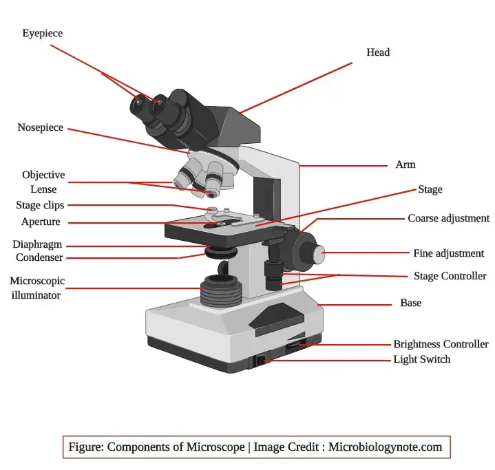
Parts of a microscope with functions and labeled diagram

Label a Microscope Worksheet by NC Middle School Resources | TpT

Draw a well labelled diagram of a microscope. - Brainly.in

Compound Microscope- Definition, Labeled Diagram, Principle ...

Microscope Parts, Types & Diagram | What is a Microscope ...

microscope drawing with label - Clip Art Library

Can someone can send me diagram of this compound microscope ...

Simple Microscope - Diagram (Parts labelled), Principle ...

Label The Microscope Parts! Diagram | Quizlet
Modified Science Diagram; Label Parts of a Microscope ...

Microscope Maintenance Tips | Science supplies, Multi step ...
Labelling A Microscope Teaching Resources | Teachers Pay Teachers
Microscope labelling 11 - Teaching resources
Post a Comment for "45 diagram of microscope with label"