43 nucleus electron micrograph labelled
Virtual EM Micrograph List | histology 021. Plasma Cell: This electron micrograph shows a typical secretory cell, a plasma cell, which secretes immunoglobulin protein. Many of the major types of cellular organelles are visible in this image. In the nucleus, areas of euchromatin and heterochromatin can easily be identified. Virtual Slide. Nucleus: Definition, Structure, Functions - Biology Learner Oct 14, 2021 · The electron micrograph and immunocytological techniques show that three distinct regions are observed in the nucleolus. These are the Fibrillar center, Dense fibrillar component, and Cortical granular components. Fibrillar Center: This pale-staining part represents the innermost region of the nucleolus, which is made up of ribosomal DNA.
[Immune electron microscope determination of the localization of ... The number of particles observed over diffuse chromatin equals to 50-80% against the label in fibroblast cytoplasm. In contrast, the label used to be absent over the E. coli nucleoid. The presence of TRS in the fibroblast nucleus may evidence in favour of a possible regulatory role of TRS in eukaryots. MeSH terms

Nucleus electron micrograph labelled
PDF Identifying Organelles from an Electron Micrograph Courtesy of Dr. Julian Thorpe - EM & FACS Lab, Biological Sciences University Of Sussex The electron micrograph displayed below illustrates many of the plant cell characteristics discussed The cell wall, large central vacuole and chloroplasts are clearly visible Also visible is the clearly defined nucleus containing chromatin Label This Transmission Electron Micrograph : TEM of chloroplast from ... Provide the labels for the electron micrograph in figure 12.8. Label the transmission electron micrograph of the nucleus. Label the transmission electron micrograph of the nucleus. Transmission electron microscopy (tem) is a microscopy technique in which a beam of electrons is transmitted through a specimen to form an image. Cell Nucleus - function, structure, and under a microscope The nucleus is a key feature that distinguishes eukaryotic cells, including all animals and plants, from prokaryotic cells (bacteria and archaea). The nucleus (plural: nuclei) stores most of the cell's genetic information in the form of DNA, although mitochondria also contain their own DNA in a very small percentage relative to the nucleus.
Nucleus electron micrograph labelled. IB Questionbank - ExamSnap 16M.2.HL.TZ0.7a: Draw a labelled diagram of a eukaryotic plant cell as seen in an electron micrograph. 16N.2.SL.TZ0.2a: The image is an electron micrograph. Determine, with a reason, whether the image is of a... 16N.3.SL.TZ0.3a: State from which organ the section was taken. Electron Micrographs of Cell Organelles | Zoology The Electron Micrograph of Nucleus: This is an electron micrograph of nucleus. (Fig. 17 & 18): (1) Nucleus was discovered by Brown (1831). (2) It is a characteristic entity of almost all eukaryotic cells except mammalian RBCs. (3) The nucleus is generally one but may also be two, four or many. Solved Label the transmission electron micrograph of the - Chegg Transcribed image text: Label the transmission electron micrograph of the cell. 0 Nucleus rences Mitochondrion Heterochromatin Peroxisome Vesicle ULAR bumit Click and drag each label into the correct category to indicate whether it pertains to the cytoplasm or the plasma membrane. ICF Contacts the ECF Made of proteins and lipids Surrounds the cell Contains lon channels Organelles Fibers ... Electron Micrograph of a Lymphocyte - Netter Images Pricing. Price for. Choose Usage Printed publication (book, brochure, journal, etc.) Trial Exhibits and Materials Slide Presentation (Non-web or authenticated login if Web) Electronic Formats Posters Tee Shirts, Novelties Student Lo-res Presentation/Poster, Thesis, Dissertation. Product Description:
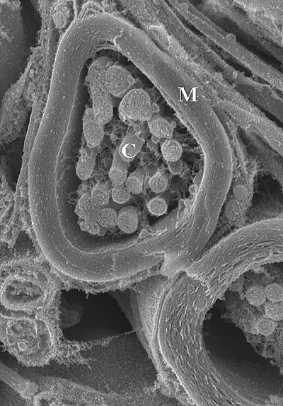
Labeling the Cell Flashcards | Quizlet Label the transmission electron micrograph of the nucleus. membrane bound organelles golgi apparatus, mitochondrion, lysosome, peroxisome, rough endoplasmic reticulum nonmembrane bound organelles ribosomes, centrosome, proteasomes cytoskeleton includes microfilaments, intermediate filaments, microtubules Identify the highlighted structures OpenLearn - Open University - A tour of the cell Figure 2 (a) A transmission electron microscope. (b) A transmission electron micrograph of a frog leukocyte (white blood cell). The nucleus and nucleolus (Section 4.3), mitochondria (Section 4.10) and Golgi apparatus (Section 4.7) can be seen. The dark area of the nucleus contains densely packed DNA. Nucleus - Electron Micrograph Slide 5 of 36 PDF Electron Micrographs (EMs) for laboratories in A215, Basic Human Anatomy There are distinct differences between cilia and microvilli to be seen in electron micrographs: - Cilia are larger (the cilium labeled C about 2.5 microns along its length is probably 5 to 10 microns long); - Cilia contain microtubules, by which they can move.
Electron micrographs of SPIO-labeled MSCs. A, Cell nucleus (N) and ... EVs can be labeled either directly using probes or indirectly by transfection of reporter genes. Optical imaging (fluorescent imaging and bioluminescent imaging), single-photon emission computed... Electron Microscopy - University of Utah Plasma cell. Normal plasma cell with prominent cytoplasmic smooth endoplasmic reticulum. Macrophage. Normal macrophage with oblong nucleus, nucleolus, and cytoplasm with a variety of inclusions. Platelets. Normal platelets. Mitochondria. Happy mitochondria within a cell. Skeletal muscle. Electron Micrographs** Electron Micrographs**. Below is a collection of electron micrographs with labelled subcellular structures that you should be able to identify. Also, be sure to observe any electron micrographs which are made available in the laboratory by the instructor. You should concentrate on the similarities in form that permit identification of the ... Electron Micrograph of a Lymphocyte Pricing. Price for. Choose Usage Printed publication (book, brochure, journal, etc.) Trial Exhibits and Materials Slide Presentation (Non-web or authenticated login if Web) Electronic Formats Posters Tee Shirts, Novelties Student Lo-res Presentation/Poster, Thesis, Dissertation. Product Description:
Kernplasma Dr.Jastrows EM-Atlas
Labeled Diagram Of Cell Membrane : Electron Micrograph The nucleus and mitochondria are two examples. Copy of labeling cell membrane labelled diagram. Some of the major parts of the plasma membrane are : Phospholipid bilayer · phospholipid bilayer ; It supports and helps maintain a cell's shape. 1)cell membrane 2)vacuole 3)nucleus 4)endoplasmic reticulum 5)mitochondria 6)golgi body.

Zellkern Dr.Jastrows EM-Atlas
Electron Micrographs Below is a collection of electron micrographs with labelled subcellular structures that you should be able to identify. Also, be sure to observe any electron ...
Gallery2 - Keele University
Plant Cell Nucleus Electron Micrograph : Cell And Organelles Dr Jastrow ... Below is a collection of electron micrographs with labelled subcellular structures that you should be able to identify. In mammals it's average diameter is about 6 an electron micrograph of a section through an animal cell nucleus (from an insect cell). In flowering plants, this condition occurs in sieve tube elements.74.

Quia - Cell Parts and Functions Flash Cards
Nucleus - The Cell: The Histology Guide - University of Leeds This picture shows an electron micrograph of a nucleus. The short white arrows are pointing to nuclear pores. Note the appearance of eu- and heterochromatin ...

The Cells and Microorganisms Webquest
Solved Please label the electron micrograph to assess your | Chegg.com Question: Please label the electron micrograph to assess your knowledge of the structure and function of a cell's nucleus nuclear pore endoplasma reticulum chromatin nucleolus nuclear envelope This problem has been solved! See the answer Show transcribed image text Expert Answer 100% (3 ratings) 1 ) Nuclear envelo … View the full answer

Search in gallery
Cell Nucleus - function, structure, and under a microscope The nucleus is a key feature that distinguishes eukaryotic cells, including all animals and plants, from prokaryotic cells (bacteria and archaea). The nucleus (plural: nuclei) stores most of the cell's genetic information in the form of DNA, although mitochondria also contain their own DNA in a very small percentage relative to the nucleus.

The Endomembrane System and Proteins | Boundless Biology
Label This Transmission Electron Micrograph : TEM of chloroplast from ... Provide the labels for the electron micrograph in figure 12.8. Label the transmission electron micrograph of the nucleus. Label the transmission electron micrograph of the nucleus. Transmission electron microscopy (tem) is a microscopy technique in which a beam of electrons is transmitted through a specimen to form an image.

#4. Cell structure and function | Biology Notes for A level
PDF Identifying Organelles from an Electron Micrograph Courtesy of Dr. Julian Thorpe - EM & FACS Lab, Biological Sciences University Of Sussex The electron micrograph displayed below illustrates many of the plant cell characteristics discussed The cell wall, large central vacuole and chloroplasts are clearly visible Also visible is the clearly defined nucleus containing chromatin
![Untitled Document [www.stolaf.edu]](https://www.stolaf.edu/people/giannini/cell/nuc/cell127.gif)
Untitled Document [www.stolaf.edu]
Post a Comment for "43 nucleus electron micrograph labelled"